
Lumps and bumps on your dogs skin can certainly be a cause for concern, but many are benign (not harmful) or are easily treated. However, malignant (cancerous) lumps and bumps can look very similar to benign skin masses, so it is always a good idea to have your veterinarian examine any new bumps. This article will discuss some of the most common causes of skin bumps in dogs and their treatment options.
Contents
- Bumps on Dogs Skin – Causes
- Fatty Tumors (Lipoma)
- Warts
- Sebaceous Cysts
- Sebaceous Adenoma
- Abscesses (Boils)
- Mast Cell Tumor
- Hygromas
- Lymphoma
- Flea Bites
- Spider and Caterpillar toxins
- Inflammatory response
- Histiocytomas
- Squamous cell carcinoma
- Skin allergies
- Diagnosis of Skin Bumps
- Treatment of skin bumps
- When to Be Concerned About Canine Skin Bumps
Bumps on Dogs Skin – Causes
Dogs can develop skin bumps ranging from little, pimple-like, raised bumps known as papules to larger ones referred to as nodules. Any dog, even a puppy, can develop these skin “masses” as veterinarians often describe them, but they tend to occur more in older dogs. They can occur on any one part of the body, or may be prevalent all over the dog’s body.
Fatty Tumors (Lipoma)
Lipomas, sometimes called fatty tumors, are some of the most common bumps found on a dog’s skin. Lipomas are round, soft, non-cancerous growths of adipose tissue (fat) that occur just under the skin and move freely when touched.. Lipomas initially present themselves as small bumps ranging between one and eight inches in diameter, but they may grow even larger over time.
Lipomas generally occur in older, overweight female dogs on the trunk (shoulders to hips) or near the tops of the legs. Breeds considered most at risk include Doberman pinschers, Labrador retrievers, miniature schnauzers, and mixed breed dogs.
Even though lipomas are benign, it’s still recommended that you have your veterinarian confirm that a suspected lipoma is indeed just a lipoma. Some lipomas, called infiltrative lipomas, will spread into the surrounding muscle and connective tissue and are considered partially malignant. Middle-aged female Doberman pinschers, Labrador retrievers, miniature schnauzers, and mixed-breed dogs are most at risk for infiltrative lipomas. Liposarcomas are rare malignant tumors that develop in older male dogs; beagles and Shetland sheepdogs are the breeds most at risk. Both infiltrative lipomas and liposarcomas look and feel very similar to the benign lipomas; your veterinarian may recommend a fine needle aspiration of the fatty tumor in order to distinguish between a benign and malignant tumor.
Ordinarily, lipomas don’t require treatment but if they cause discomfort to your dog, or if they have grown large enough to interfere with normal body movement, they can be removed surgically. Surgical removal of lipomas, infiltrative lipomas, and liposarcomas is considered curative if all of the abnormal growth is removed. Because the abnormal lipoma cells tend to mix in with normal fat cells, your veterinarian may recommend putting your dog on a diet for several weeks before surgery in order to make it easier to determine where the tumor ends and normal fat tissue begins.
Warts
Also known as cutaneous papillomas, warts are benign, light-colored, hard bumps that resemble cauliflower in shape. In young dogs, they are caused by a virus and appear in and around the mouth (on the lips, tongue, etc.) and also around the eyes; multiple warts will appear at the same time. If the inside of the mouth becomes severely affected, it is possible for the warts to interfere with chewing and swallowing. However, the virus is usually self-limiting, and will resolve on their own over the course of weeks to months. A drug called azithromycin has been shown to speed up the resolution of the warts.
In older dogs, they can occur anywhere on the body, usually singly, and have not been definitively linked to viral infections. They can be confused with skin tags. Cocker spaniels and Kerry blue terriers seem to be predisposed to these types of warts.
Warts typically heal on their own but surgery may be recommended to remove irritated or frequently bleeding warts. Warts that occur on the eyelids may also be a candidate for surgical removal, since they could possibly irritate the eye. Rarely, viral papillomas may progress to invasive squamous cell carcinomas, so it is important for you and your veterinarian to closely monitor such bumps.
Sebaceous Cysts
A sebaceous cyst is a skin lump that occurs when the sebaceous (oil) glands in a dog’s skin become blocked. It may eventually burst and discharge a white, cheese-like substance. As tempting as it may be, you should not attempt to squeeze out the contents of a sebaceous cyst because the contents may be forced deeper into the skin and cause a localized inflammatory reaction and/or an infection.
Sebaceous Adenoma
Sebaceous adenomas are shiny skin-colored masses with small nipple-like projections; the masses are usually a quarter of an inch to an inch in diameter. These benign tumors arise from the sebaceous glands of the dog’s skin, and often appear in multiples anywhere on the dog’s skin, but are usually found on the dog’s head.The breeds most likely to develop these tumors include coonhounds, English cocker spaniels, cocker spaniels, huskies, samoyeds, and Alaskan malamutes. Treatment usually consists of monitoring the masses unless they grow, change color, or become irritating to the dog. In that case, surgical removal will be recommended and will cure the adenoma at that particular site, but more adenomas may appear in other places as the dog ages.
Abscesses (Boils)
Abscesses, an accumulation of pus under the skin, are yet another likely cause of bumps under a dog’s skin. An abscess usually appears suddenly as a painful swelling that is warm to the touch and may be either firm or moveable and compressible. The abscess may be large or small, and may cause redness in the surrounding skin. Dogs with abscesses usually have a fever, be lethargic, and not eating well. Some abscesses will rupture on their own and drain a foul-smelling fluid-like material. .
Abscesses are usually caused by some sort of penetrating wound, such as an animal bite or other physical injury such as being stuck by a stick or a grass seed. Sites of former infections can also serve as a source for an abscess, especially in the anal glands. If a penetrating wound occurs and the wound closes up without allowing enough drainage of the introduced bacteria, an abscess will often form. Treatment of abscesses usually involves opening, draining, and flushing the pocket of pus. If a foreign object such as a stick or grasses caused the abscess, it’s important to make sure that is removed as well, otherwise new abscesses will form. Treatment with antibiotics and pain medications will follow drainage of the abscess. It is important to complete the full course of antibiotics to prevent recurrence of the abscess.
Mast Cell Tumor
Mast cell tumors are the most common malignant canine skin cancer. These tumors may be seen in dogs of any age, but are most common in older dogs (8-10 years of age), and may develop anywhere on the body, but are most commonly found on the limbs and the underside of the thorax and abdomen. The tumors vary greatly in size and appear as raised, nodular masses that can feel firm to soft on palpation. Several breeds appear to be predisposed, including pugs and boxers, in which multiple mast cell tumors form, and also Rhodesian ridgebacks and Boston terriers.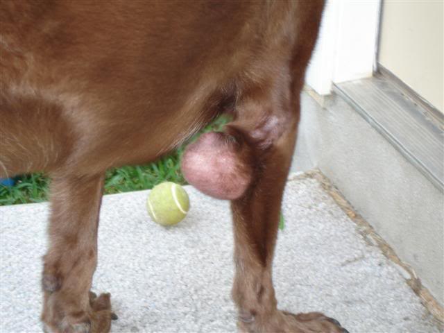
Diagnosis of a mast cell tumor is done with a fine needle aspirate, which is inserting a needle into the mass, suctioning out some of the cells, and examining the cells under a microscope. Once diagnosed, surgical removal is recommended. The surgeon will attempt to remove as much of the surrounding tissue as possible, since these tumors have a halo of abnormal cells around them that are impossible to distinguish from normal tissue by look or feel. Prognosis depends on the grade of the tumor as determined by a veterinary pathologist; survival time could be as little as four months, or as much as two years or more. A combination of chemotherapy and radiation therapy is usually recommended for the best prognosis.
Hygromas
Hygromas are soft, painless, fluid-filled sacs that form over bony prominences such as the elbow, or over pressure points such as over the hip joint. In younger dogs, when hygromas appear, they are thought to be caused by trauma. In older large breed dogs, hygromas usually form in response to impaired movement and excessive time spent lying down on hard surfaces; formation over the elbows is particularly common, especially in dogs with elbow arthritis.
If caught early while they are still small, treatment consists of draining of the fluid by a veterinarian, followed by better and thicker bedding for the dog. Small hygromas can also be treated with laser therapy, which encourages healing of the site. For chronic hygromas, surgical drainage of the hygroma is recommended, followed by flushing and placement of Penrose drains (a soft rubbery tube that is placed in the wound and allowed to exit the skin; this permits continued drainage of the site for several days after surgery). Hygromas that have become ulcerated require a more aggressive treatment, up to and including skin grafts and antibiotics.
Lymphoma
Lymphoma is a blood cancer characterized by uncontrolled growth in the numbers of lymphocytes, a type of white blood cell. These cells clump together in the lymph nodes, causing swelling of those nodes. Lymph nodes can be found near the base of the lower jaw (mandible), in front of the shoulder blades, in the “armpits”, in the groin, and on the back of the legs between the knees and the ankles. Enlarged lymph nodes can also be caused by localized infection or inflammation, so it is important to have cells from the lymph node examined under a microscope for diagnosis of lymphoma. Lymphoma can be of B-cell or T-cell type; B-cell lymphoma is more common and has a better long-term prognosis.
Internal lymph nodes in the chest and abdomen can also be affected, as well as the spleen and liver. Treatment can be palliative (improving the quality of life) with oral steroids such as prednisone; average survival time with palliative care is usually 3-4 months. A combination of oral and intravenous chemotherapy with four different drugs is considered the gold standard of care in B-cell lymphoma and usually results in clinical remission in 90% of cases; survival time with this treatment is usually 12-14 months before relapse. Newer treatments showing good response include half-body or whole-body radiation therapy, bone marrow transplant, and the infusion of antibodies or the use of vaccines.
Flea Bites
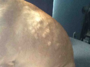
Also referred to as flea bite sensitivity, flea allergy dermatitis can also manifest itself in skin bumps. These tiny red bumps are usually found on the dog’s stomach and in the groin area. The condition occurs when the dog’s immune system reacts to flea saliva. In addition to bumps, your canine may also show symptoms such as redness, hair loss, crusty or scaly skin, and intense itching.
For diagnosis, your vet will start by checking your dog’s skin for the presence of fleas after which he or she may follow up with intradermal testing. Treatment revolves around flea control with insecticides such as permethrin. [Capstar] is an oral medication that can be used to kill fleas on your dog within 30 minutes. Your vet is also likely to prescribe steroids to control the swelling, antihistamines to curb the itching, and recommend the use of a flea and tick preventative such as the [Seresto collar], topicals such as [K9 Advantix II], [Vectra 3D], or [Frontline Plus], or prescribe an oral medication such as NexGard, Simparica, or Bravecto.
Spider and Caterpillar toxins
Dogs can be exposed to spider and caterpillar toxins as a result of spider bites or contact with caterpillars respectively, leading to a condition medically referred to as eosinophilic folliculitis. This is marked by swelling of the affected area, usually the nose in dogs, alongside other symptoms such as redness, pain, and eventually draining ulcers.
Treatment of the condition involves the administration of corticosteroids and dressing the wounds. There is always the risk of permanent hair loss and scarring.
Inflammatory response
Some bumps on a dog’s skin are in reaction to something that causes localized inflammation, such as a bee sting, tick bite, or a reaction to an injection. Bee stings are usually treated with intravenous steroids and an injection of antihistamine, especially if the sting is around the mouth of the dog, causing swelling of the muzzle and potentially making it difficult for the dog to breathe. Bumps from a tick bite will usually resolve on their own after a few days, but careful monitoring of the location is always a good idea. Watch for increased irritation, redness, or swelling. A reaction to an injection such as a vaccine would be expected to dissipate after a day or two, but an excessive reaction may also be treated with steroids and antihistamines, and some veterinarians will pre-treat a dog with known vaccine reactions before administering the vaccine.
Histiocytomas
Histiocytomas are benign tumors of mainly young dogs. The tumors are solitary, raised, hairless bumps that develop rapidly and may become ulcerated. They are most commonly found on the head, ears, and limbs of the dog. Breeds most at risk include greyhounds, English bulldogs, Scottish terriers, boxers, and Boston terriers. Histiocytomas usually resolve on their own within 2-3 months and only require surgical intervention if they become bothersome for the dog.
Squamous cell carcinoma
Squamous cell carcinomas are the most commonly diagnosed epithelial cancers arising from the skin. They are firm, nodular masses that may be raised and/or ulcerated, with older bloodhounds, basset hounds, and standard poodles at the greatest risk. While the cause for most squamous cell carcinomas is unknown, prolonged sun exposure is a risk factor for some of these tumors. When sun injury is the cause, patients are often short-haired dogs with white skin, such as dalmatians, bull terriers, pit bulls, and beagles. Surgical removal of the mass is the treatment of choice.
Skin allergies
Environmental or food allergies may present as small, reddened, itchy bumps on the skin. Other symptoms of allergies include darkening of the skin, hair loss, fur staining (as a result of repeated licking), frequent shaking of the head, scratching at the ears, and ear infections (which may then result in foul smelling discharge).
Excessive scratching warrants the attention of your veterinarian who will do a physical examination of your dog and then recommend appropriate tests to identify the underlying causative factor. Among the tests they may consider are ear cytology, skin cytology or biopsy, allergy testing of the skin or blood, or food elimination diets. The veterinarian may also ask if you have recently changed your dog’s food or diet.
Your veterinarian will also check your dog for other conditions known to cause itching in dogs including flea and other parasitic infestations, bacterial infections, fungal infections, and systemic diseases.
Once your veterinarian has ascertained the underlying factors for the itchy bumps, they will then prescribe the appropriate medications. Among the treatment options are:
- Immunotherapy: Involves exposing the dog to gradually increased amounts of the allergen to help desensitize its immune system.
- Steroids
- Antihistamines: This helps to ease the itching associated with dog allergy bumps.
- Parasitic control (when flea or other parasites are the underlying factors for the bumps).
- Diet change – your vet may recommend switching to a new dog food with a new protein source or even help you prepare a diet for your dog.
In addition to the above treatment options, you may also want to consider the following tips:
- Change air conditioning filters often
- Wash your dog’s bedding at least once every week with hot water
- Vacuum the house often
- Use dust mite barriers to cover your dog’s beddings.
- Avoid walking your dog outdoors when pollen levels are at their peak
- Wash your dog’s food bowls often with warm, soapy water
- Keep the dog off the lawn after it has been mowed
Other causes of dog skin bumps include:
-
Folliculitis – Infected hair follicle.
-
Hematomas (blood blisters – These are caused by trauma and usually resolve on their own, but surgical drainage may be required.
-
Canine acne (yes dogs can also get acne)
-
Hookworms – causes red bumps on a dog’s feet which is typically accompanied by itching and abnormally growing nails.
-
Skin tags – Soft tiny growths that are usually a few millimeters in length but could grow to several inches and are usually attached to the body by a thin stalk Generally no treatment is necessary.
-
Other types of skin tumors
Diagnosis of Skin Bumps
Your veterinarian may be able to recognize some bumps, e.g., lipomas or sebaceous cysts by observation. However, fine needle aspiration of the bump is always a good idea since some malignant bumps resemble benign

masses. Fine needle aspiration entails inserting a needle attached to a syringe into the growth to extract a few cells for examination under the microscope.
If your veterinarian suspects something more serious like cancer, they will take a small tissue sample from the affected area and send it out for biopsy. For cancerous bumps, surgical removal, chemotherapy, and radiotherapy may be considered.
Your veterinarian will also want a brief history of your dog and the mass including:
- The onset of the symptoms
- Whether the lump appeared suddenly
- Whether the shape or size of the lump has changed
- Whether the condition has had any impact on your dog’s appetite or energy level
- If you have recently introduced your dog to a new food (which may have triggered allergic reactions)
- If your dog has recently had a parasitic infestation especially fleas
Treatment of skin bumps
Surgical removal is the most common way to get rid of dog skin bumps but it is not always required. Some conditions such as histiocytomas and sebaceous cysts may resolve on their own without treatment. Your veterinarian will make recommendations as to the best course of action.
When to Be Concerned About Canine Skin Bumps
You should talk to your vet immediately about a bump if:
- You are not sure what the underlying cause is
- It bothers you or your pet
- It grows rapidly
- It occurs on or around the eyelids where it may interfere with vision or be difficult to remove later on when it has grown larger. The latter applies to bumps that appear on the paws and face as well.
- It is reddened, swollen, or oozes blood or pus
- Causes pain to your canine friend