IN THIS POST
As dogs age, they are more prone to getting lumps and bumps. One of the common types of bumps are lipomas, also sometimes called fatty tumors. While the word “tumors” sounds a bit scary, tumors are simply growths of abnormal tissue. Benign tumors are usually localized, and do not spread to other parts of the body (metastasize), whereas malignant tumors invade local tissue and can spread to other parts of the body. As with most fatty tumors on dogs, lipomas don’t have a clear cause; genetics, hormones, environment, and diet all can have some influence on their development. In this article, we’ll talk about lipomas and similar tumors, their diagnosis, and their treatment.
Fatty Tumors On Dogs
Lipomas
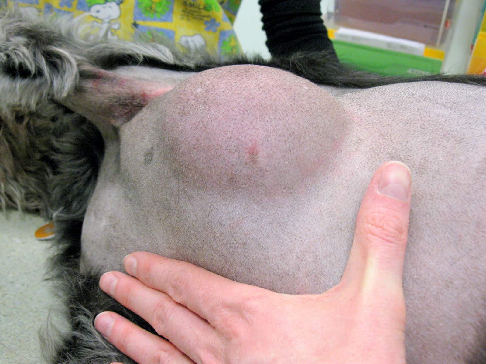
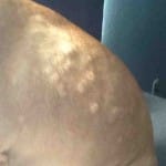
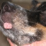
Lipomas are benign (non-cancerous) tumors made up of adipose (fat) tissue. They usually appear as discrete, soft, lumpy masses just under the surface of the skin and most will move freely when manipulated. Lipomas are generally found on the trunk (shoulders to hips) and near the top of the legs. However, in rare cases, they can also form in the abdomen, where a layer of adipose tissue cushions the abdominal organs. Older, obese female dogs are more likely to develop them. Doberman pinschers, Labrador retrievers, miniature schnauzers, cocker spaniels, Weimaraners, dachshunds, and small terriers are the breeds most at risk of developing lipomas.
Most veterinarians will feel fairly confident in diagnosing a lipoma based on the age and breed of the dog, location of the mass, and the way the mass feels while being palpated. However, it is important to get a definitive diagnosis because 10-15% of mast cell tumors, which are malignant tumors, closely resemble lipomas by sight and feel. Since mast cell tumors have a better prognosis the earlier they are found, having every lump and bump checked out will aid in finding any possible malignant tumors that much sooner.
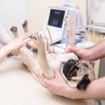
Diagnosis of many tumors can be achieved via examination of cells collected from the mass with a procedure called fine needle aspiration (FNA). In this procedure, a needle attached to a syringe is inserted into the mass. The veterinarian will move the needle around to a few different places within the mass while pulling back on the syringe. This creates negative pressure within the syringe, drawing cells from the mass into the needle and syringe. The contents of the syringe are then “shot” onto several microscope slides by drawing back on the syringe and then quickly pressing the plunger. The slides are then stained and examined under a microscope.
 Since lipomas are composed of adipose (fat) tissue, visualization of this type of tissue on the slide is fairly diagnostic. Adipose tissue has very characteristic large, empty-looking cells, and often the material on the slide itself has a greasy texture to it.
Many masses may have more than one type of tissue present, and so one drawback to fine needle aspiration is that it may be possible to miss sampling some cells, leading to a missed diagnosis. Because fine needle aspirates are not 100% accurate or diagnostic, your veterinarian should ask you to monitor the mass closely for any changes in size, appearance, or texture. If those occur, resampling of the mass with fine needle aspiration should be done at a minimum; your veterinarian may recommend a biopsy or complete removal of the tumor instead.
Many veterinarians will usually recommend a wait-and-see approach to lipomas. If the mass remains relatively small, slow-growing, and in a location that doesn’t interfere with the dog’s movement, that may not be an unreasonable approach. Of course, if anything changes significantly, your dog should return to the veterinarian for re-evaluation as soon as possible.

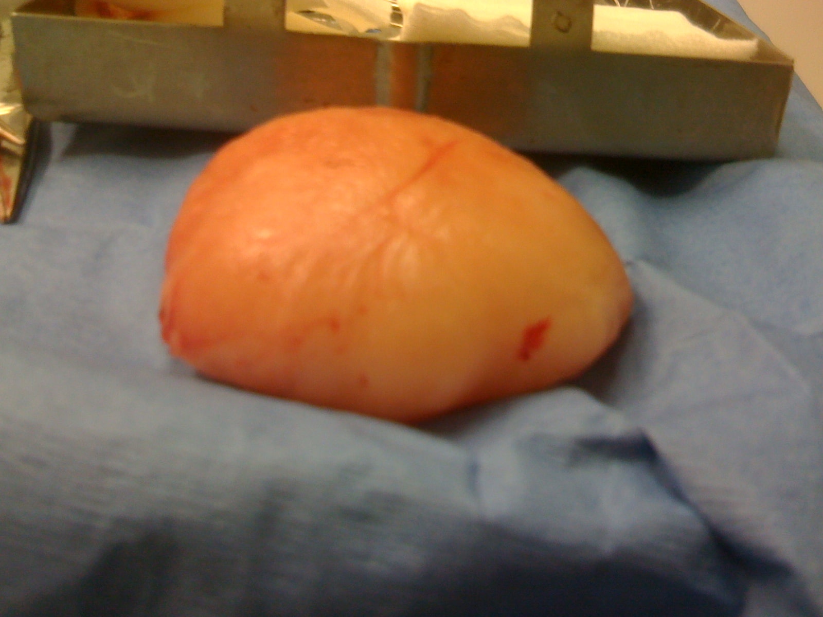
Another option is to forego the wait-and-see approach and proceed directly to surgical removal. The advantage to going directly to surgery is that the surgery is much easier while the mass is smaller. Many lipomas merge with the healthy fat tissue next to them, which makes it difficult for the surgeon to determine the edges of the tumor and therefore uncertain if they removed all of it. Some surgeons may recommend a weight-loss regimen for the dog starting several weeks before surgery; this may make it easier for the surgeon to visualize the edges of the tumor, ensuring complete removal.
Another advantage of proceeding directly to surgery is that benign lipomas may be indistinguishable from the more malignant infiltrative lipomas and liposarcomas, so removing the tumor early on will improve the prognosis, should the tumor turn out to be one of those types of tumors. Infiltrative lipomas and liposarcomas are discussed in more detail later in this article.
Surgical removal will also be more strongly recommended for large masses that interfere with the dog’s movement, cause discomfort, or interfere with normal bodily functions.
Other treatment options for lipomas such as using steroid injections, liposuction, and laser therapy have been reported in the veterinary literature. The treatments had varying degrees of success and the number of patients in the studies were fairly small, so the jury is still out on adopting any of those options for treatment at this time.
There is a rare variant of lipomas found in dachshunds, called diffuse lipomatosis, in which nearly the entire skin is affected, resulting in rolls of fat around the neck and trunk. Unfortunately, there is no treatment for this type of lipoma, though some veterinarians recommend surgical removal of any areas of tumor tissue that interferes with the dog’s quality of life. However, this is only a temporary fix as the tumor tissue will regrow.
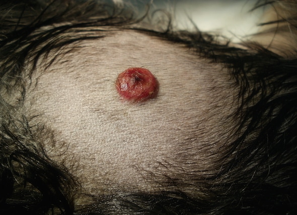
Infiltrative Lipomas
Infiltrative lipomas are very locally invasive tumors that arise from the fat cells and invade deep tissue such as muscle, nerves, and bone. However, they rarely spread to other sites (metastasize), and so are considered only partially malignant. The tumors appear to be soft, lumpy swellings in the fat layer under the skin, usually on the chest and legs. They are very rare in dogs, but are most common in middle-aged female doberman pinschers, Labrador retrievers, and miniature schnauzers.
Diagnosis of these tumors can be difficult since results of a fine needle aspirate often appear the same as the more benign lipomas. The diagnosis comes from advanced imaging (CT or MRI), evidence of deep tissue involvement at the time of surgery for lipoma removal, or from pathology results of the removed tumor. Thus, it may be prudent to consider surgical removal of any lipomas on dogs that have been shown to be most at risk for infiltrative lipomas.
Whenever possible due to the location of the tumor, surgical removal is recommended. The surgeon will remove as much of the tumor as possible (debulk the tumor), but due to the infiltrative nature of the tumor, it is nearly impossible to remove all of it. Surgery alone results in a 40% recurrence rate, and half of those cases recur within 8 months. Dogs that are treated with surgery alone lived about 18 months after diagnosis. The exception is if the tumor is on a leg — amputation of the entire limb would likely remove the entire tumor and be considered curative.
Radiation therapy is usually recommended in conjunction with surgical debulking of the tumor. Radiation therapy consists of targeted doses of radiation daily for 15-18 treatments. The dogs are under general anesthesia for the 15-30 minutes of treatment, since they need to be absolutely still for correct targeting of the radiation. Side effects are generally mild, and include redness, irritation, and loss of fur in the area being treated. In one published study, 50% of dogs that had surgery followed by radiation therapy survived at least 40 months. In general, over 90% of dogs that have a full course of radiation therapy after surgery are disease free for at least a year.
While it is possible to just do radiation therapy without the surgery, it’s generally not recommended. Radiation therapy is more effective when smaller amounts of tumor tissue is present, so debulking the tumor surgically first provides the best results. Chemotherapy is generally not effective for localized tumors such as infiltrative lipomas, and as such, is not usually recommended. In rare cases, a low-dose chemotherapy protocol designed to reduce the formation of new blood vessels may be used to prevent tumor recurrence by limiting new blood supply to the area of the tumor.
Liposarcomas
Liposarcomas are rare malignant tumors of the adipose tissue underneath the skin. They are solitary masses without a clearly defined border. They usually develop on the chest or legs and can be either soft or firm to the touch. Liposarcomas are locally invasive, and may spread (metastasize) to the lungs, liver, spleen, or bones, though metastasis is not as likely with liposarcomas as it is with other cancers. Older male Shetland sheepdogs and beagles are most at risk for liposarcomas.
Unfortunately, fine needle aspiration of a malignant liposarcoma usually yields cells that look like a benign lipoma. Thus a biopsy of the mass is usually needed to make a diagnosis; two different types of biopsies can be performed. With a tissue biopsy, your dog is administered a local anesthetic, and a small piece of the lump is removed and sent to the laboratory for analysis. While a definitive diagnosis is very likely with this approach, a drawback is that if the pathology results indicate a malignant growth, another procedure will have to be performed to remove the growth.
An excisional biopsy is performed while your dog is under general anesthesia and involves the removal of the entire mass and as much of the surrounding tissue as possible to ensure that all of the tumor has been removed. This approach will yield a definitive diagnosis, and if it is a malignant growth, the pathologist will also comment on whether they believe all the tumor cells have been removed from the surgical site. This is done by evaluating the margins, or edges, of the removed tumor, looking for tumor cells among the normal tissue.
A recent study showed that a liposarcoma diagnosis can also be made using a CT scan of the mass. A CT scan is basically a 3D X-ray of the area of interest. Because the dog cannot move during the time it takes to run the scan (between 20 and 60 minutes), general anesthesia is used to keep the dog still. Many specialty and referral veterinary practices have a CT scanner on site.
Similar to infiltrative lipomas, surgical removal or debulking of the mass is the treatment of choice. Dogs who are diagnosed with liposarcoma by excisional biopsy but do not have the mass removed live an average of 183 days, or about six months. If the surgeon is unable to remove all of the tumor because of its location, survival time averages 649 days, or about a year and a half. If the surgeon was able to remove all of the tumor (called having “clean margins”), the average survival time increases to 1188 days, or over three years.
Some studies are being done combining radiation therapy with surgical debulking of liposarcomas, particularly in cases where the surgeon was unable to achieve clean margins. Final results are still pending, but preliminary work appears promising.
Final Thoughts
While jumping straight to surgical removal for presumed lipomas is probably not realistic for most owners, and may not be in the best interests of every dog, it is certainly important to make sure to have your veterinarian check lumps and bumps with a fine needle aspirate. Malignant mast cell tumors can look similar to lipomas in appearance, and some veterinarians have been caught off guard with assuming a dog has yet another lipoma only to find out differently when a fine needle aspirate was performed. If your dog is in one of the risk groups for infiltrative lipoma or liposarcoma, then keep in mind that a fine needle aspirate is likely not going to diagnose those more serious tumors, and at the very least, a tissue biopsy should be performed of any new masses. With careful monitoring of your dog’s lumps and bumps, you and your veterinarian can work together to keep your dog healthy for as long as possible.
Related posts:
 Lipomas in Dogs, Infiltrative, Causes, Removal and Pictures
 Bumps on Dogs Skin, Back, Head, Chin, Face and Nose – Treatment Options
 Bumps on Dog Lips: A Clinical Guide to Oral Growths
 Hard Swollen Stomach in Dogs, Bloat, Causes & Remedies
 Warts on Dogs: Symptoms, Causes, Treatment, and Prevention
 Bump on Dog Eyelid, Cyst, Stye, Growth and Tumors